
Fig. 3.
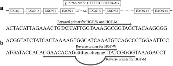
Alternative splicing of bovine HGF gene. a The genomic location of splicing fragment (gray region) in HGF-M was compared to HGF-W. Boxes show the exons and lines represents introns. Dashed line indicating the partial genomic structures are not listed. b Positions of primers for detecting the HGF-W and HGF-M mRNA expression level. Lower cases show the splicing fragment sequence. The bow line indicates that the reverse primer for HGF-M skips the splicing fragment