Fig. 1.

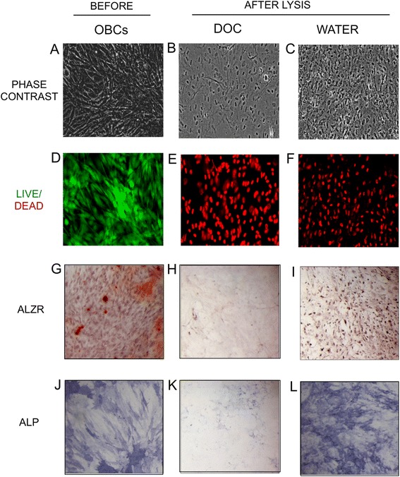
Lysis of osteoblastic cells. MSC cultures were osteogenically differentiated for 15 days, lysed with DOC or water, and examined microscopically. a-c Phase contrast images of residual matrix after lysis. d-f Fluorescent live (green)/dead (red) merged images. g-i AlzR staining of matrix calcification. j-l blue staining of ALP enzymatic activity. (a, d, g, j) OBCs before lysis. (b, e, h, k) OBCs after DOC lysis. (c, f, i, l) OBCs after water lysis. Microscopy images taken at 10×. MSC mesenchymal stem cells, AlzR Alizarin Red S, ALP alkaline phosphatase, OBCs osteoblastic cells
