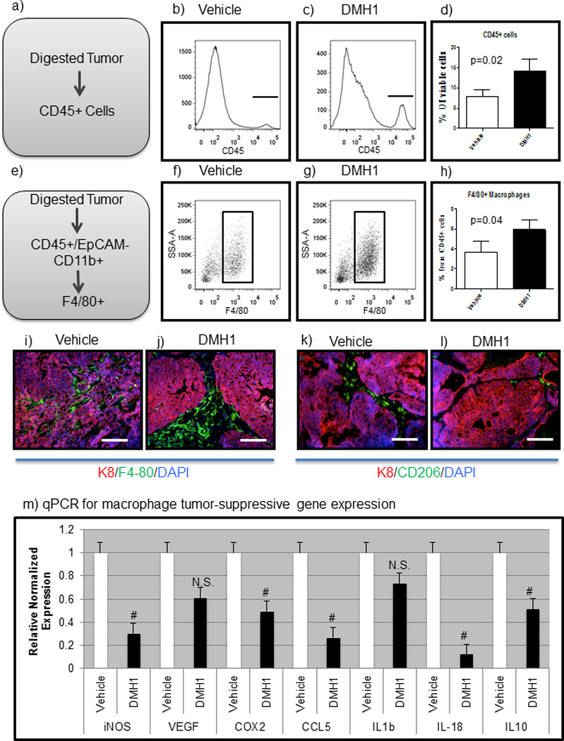
Figure 6. DMH1 inhibition for BMP signaling alters the immune response in primary tumors.

a-c) Primary tumors were digested and stained for viable CD45+ cells. d) DMH1 treated tumors demonstrated significantly increased immune cells than vehicle controls. e–g) Macrophage cells were significantly increased in DMH1 tumors as compared to vehicle controls. i–j) IF Staining for F4/80 macrophages (green) were identified within the tumor center as compared to DMH1 treated tumors which were located mostly peri-turmorally (j). k–l) Tumor promoting macrophages (CD206-green) were more abundant in vehicle treated tumors than DMH1 treated. m) FACS sorting for F4/80+ macrophages revealed distinct polarization in DMH1 tumors to be less tumor promoting than vehicle isolated macrophages with significant reductions in inflammatory cytokines. Microscope scale bars = 100µM. # Indicate statistically significant by students T-test. Error bars for qPCR and flow cytometry indicate SEM.
