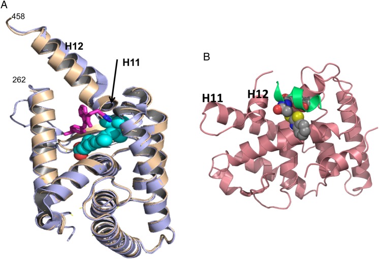
Figure 4.
Novel binding regions in RXRα (A) K-8008 binds to a novel binding region: the K-8008 binding region is away from the 9-cis-RA binding area and located on the surface of monomeric RXRα. It shows the superposition of the monomer of RXRα-LBD/K-8008 complex structure (brown) and the apo protein structure (purple, from PDB entry 1G1U). K-8008 is shown as sticks (carbon in magenta and nitrogen in blue). The classic ligand-binding site is indicated by a VDW ball model of 9-cis-RA (in cyan/red) taken from a superimposed 1FBY of PDB. (B) The proposed binding region for compound 23. The compound 23-binding region overlaps with the coactivator-binding region. Here, compound 23 was docked to the structure 3FUG (in pink) of PDB and the docked conformation (in VDW balls) was displayed with the coactivator peptide (in green) in the structure of 3FUG.

