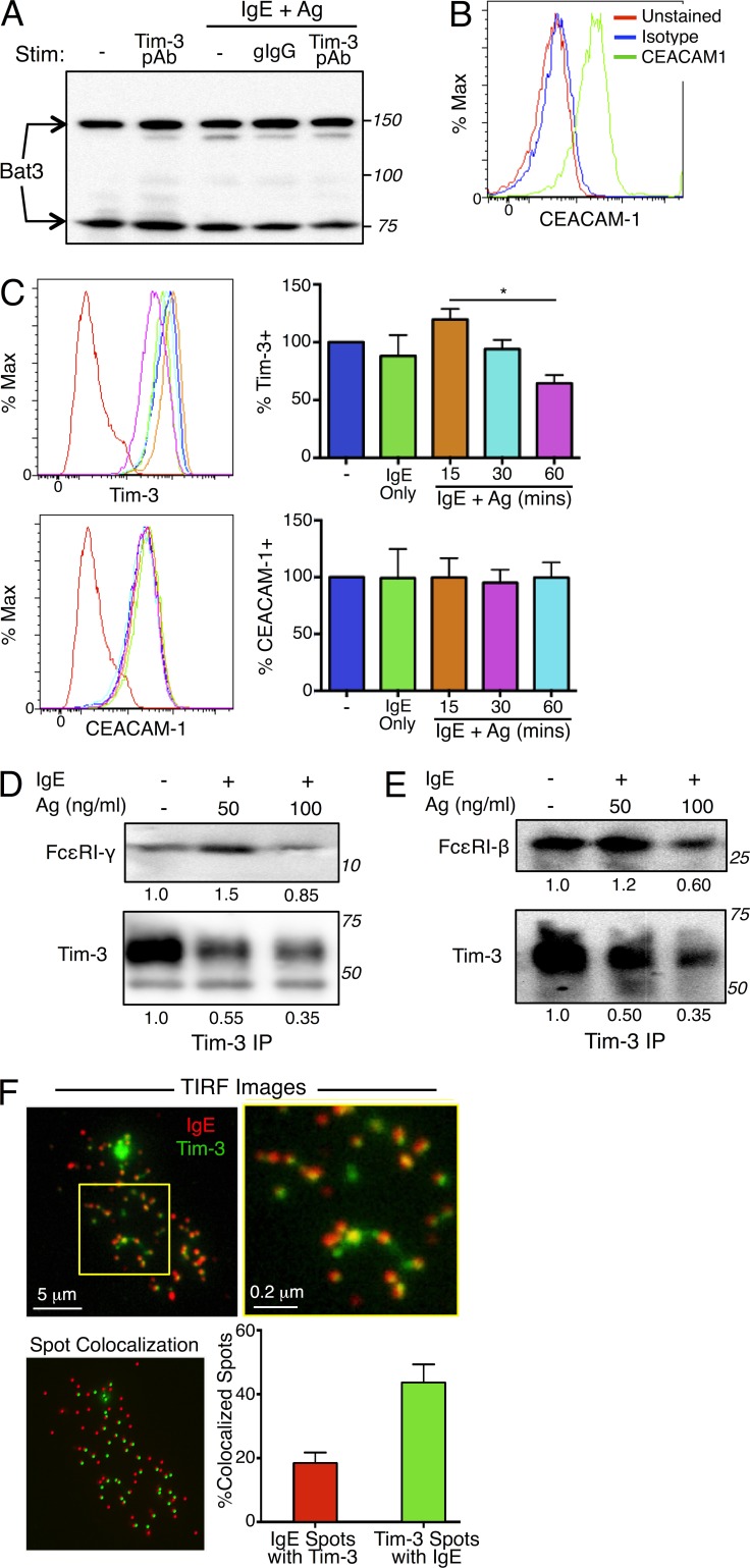
Figure 6.
Tim-3 associates with subunits of FcεRI and is comodulated with FcεRI after stimulation with Ag and IgE. (A–C) Tim-3–interacting proteins Bat3 and CEACAM1 are expressed in mast cells. BMMCs were stimulated with IgE/Ag in the presence of isotype control or Tim-3 pAb. Bat3 expression was determined by Western blotting (A). CEACAM1 expression was determined by flow cytometry in the resting state (B) or after stimulation with IgE/Ag (C); Tim-3 modulation was also assessed by flow cytometry. (D and E) Tim-3 IPs were analyzed for the presence of FcεRI γ chain (D; top) or β chain (E; top), or Tim-3 (bottom). (F) BMMCs were transfected with Tim3-mYFP, sensitized with IgE-Alexa Fluor 647 for 1 h, and settled onto poly-D-lysine–treated glass-bottom dishes coated with 1 µg/ml DNP32-HSA. Cells were fixed with 2% PFA after 1 h, and TIRF images were collected. Results are representative of three independent experiments (A–C) or two independent experiments (D–E). *, P < 0.05. The percentage of colocalized spots was derived from the average of 21 cells both labeled with anti-IgE-Alexa 647 and expressing Tim3-mYFP, in three independent experiments.

