
Figure 1.
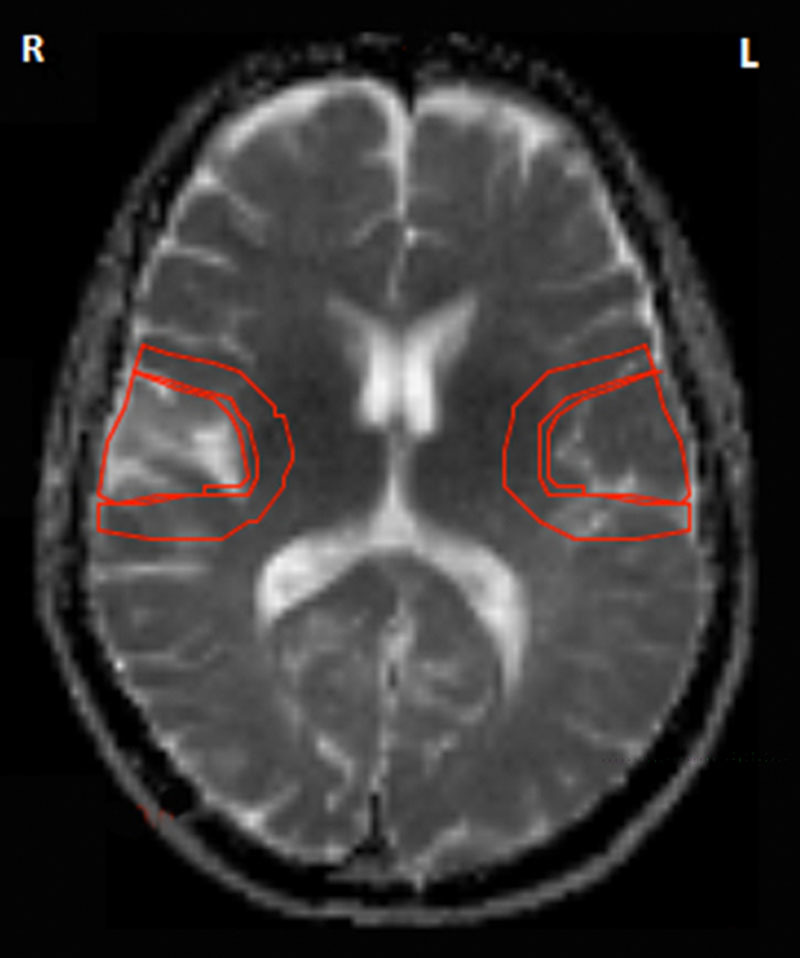
An example of stroke-specific regions of interest showing an area of ischemic infarct (right temporal region) with peri-infarct region and the mirror regions in the contralateral hemisphere. There is no evidence of hemorrhagic transformation. The representative axial image is based on an magnetic resonance apparent diffusion coefficient image at the level of the internal capsule.