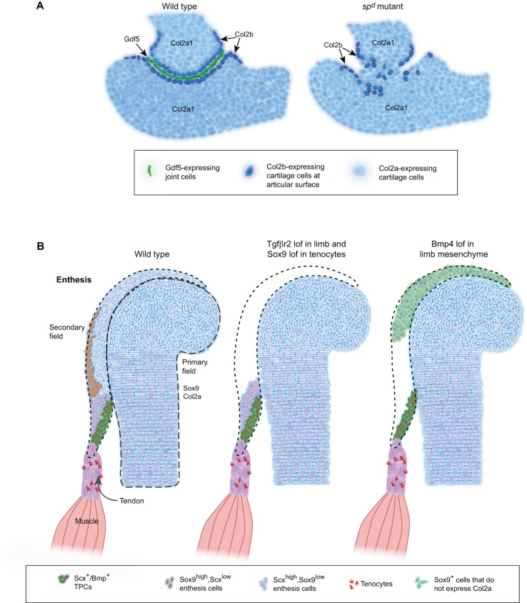
Fig. 6.
ECM functions at tendon-bone attachments. (A) Diagrams illustrating changes in cartilage at the developing humero-ulnar joint of the mouse forelimb in wild-type embryos (left) and in splotch delayed (spd, Pax3) mutants (right), which lack muscles. Proliferating chondrocytes express Col2a1 (light blue), whereas cells forming at the edges of the joint express Col2b (dark blue), and cells in the joint interzone secrete Gdf5 (green) into the joint region. The loss of muscles in spd mutants leads to loss of Gdf5 expression, disorganized Col2b+ interzone cells and joint fusion. (B) Diagrams illustrating changes in cartilage and tenocytes at a developing eminence. The primary field contains cells that form chondrocytes within the developing bone, whereas the secondary field consists of Sox9-positive progenitor cells that lie outside of the primary field. In wild-type embryos, three different subsets of Scx-expressing cells at a muscle insertion site of a developing long bone are found: Sox9+/Scx+, Scx+ or Scx+/Bmp4+. Loss of Tgfβr2 in limb mesenchyme or of Sox9 in tenocytes leads to a loss of the Sox9/Scx co-expressing and Sox9-expressing population in the secondary field, but not other tenocytes. Loss of Bmp4 signaling leads to a loss of both Sox9+/Scx+ and Scx+ populations in the secondary field. Dotted lines outline primary field. Dashed lines outline secondary field. lof, loss of function.

