Fig. 1.
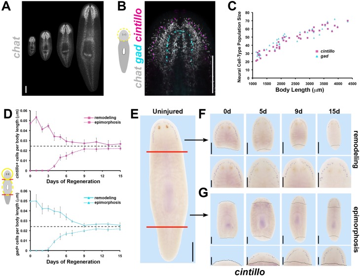
Planarian brain:body proportion is restored through regeneration by either increasing or decreasing brain cell number as necessary. (A) FISH detecting chat expression in intact animals 2-8 mm in length. (B) Triple FISH detects chemosensory neurons (expressing cintillo, magenta), GABAergic neurons (expressing gad, cyan) and cholinergic neurons (expressing chat, gray). (C) Numbers of neuronal subpopulations (cintillo+, magenta; gad+, cyan) from differently sized uninjured animals plotted against body length. (D) Average numbers of cintillo+ neurons (top, magenta) and gad+ neurons (bottom, cyan) normalized to animal length during epimorphic regeneration of a new brain (dotted lines) or remodeling of the pre-existing brain (solid lines; averages of n≥5 samples; bars, s.d.) measured by FISH. Black dashed lines in D indicate interpolated brain proportion based on analysis of intact brain proportions (see Materials and Methods). (E-G) WISH showing cintillo expression in intact animals (E), head fragments remodeling a pre-existing brain (F) and trunk fragments regenerating a new brain through epimorphosis (G). (F,G) Black dashed lines indicate amputation plane; bottom panels show higher magnification view of top panels. Scale bars: 300 µm in A,B,E and F,G top panels; 150 µm in F,G bottom panels. Anterior, top. d, day.

