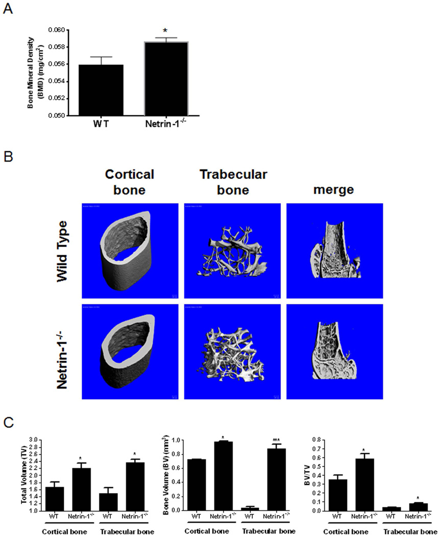
Fig. 4.
Morphometric examination of long bones in 5-month-old WT and Netrin-1−/−mice. (A) Whole-body dual X-ray absorptiometry (DXA) scanning to assess the bone mineral density (BMD) (gm/cm2) of the whole skeletons of Netrin-1−/− and wild-type (WT) mice (n = 9 each). (B) Representative high-resolution micro-CT images. Three-dimensional images of the reconstruction of the femurs revealed increased bone mass in Netrin-1−/− mice compared with their WT littermates (n = 5 each). (C) Digital morphometric analysis of micro-CT images from WT and Netrin-1+/− mice. All data are expressed as means ± SEM. ***p < 0.001, *p < 0.05 related to WT (Student’s t test or ANOVA).

