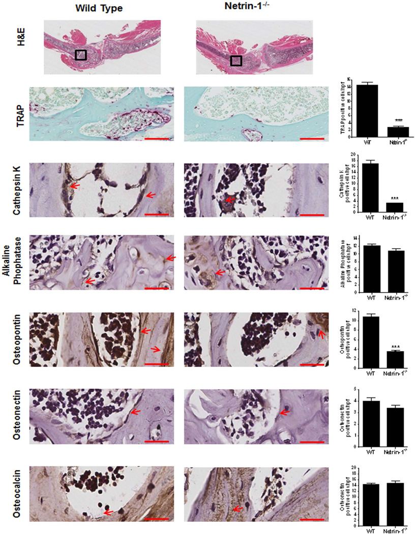
Fig. 5.
Histological examination of long bone from WT and Netrin-1−/− mice. Long bones (femur and tibias) were stained with hematoxylin and eosin to determine morphology. Representative histologic sections obtained from the femurs of WT and Netrin-1−/− mice, stained for TRAP and cathepsin K as markers of osteoclasts, alkaline phosphatase as a marker of osteoblasts. Osteopontin, osteonectin, and osteocalcin were counterstained with hematoxylin. Quantification of the number of osteoclasts/hpf (high-power field) was performed by counting positive cells in five different images for each of 3 mice. All images are taken with the same magnification. Scale bar = 50 µm. Data represent means ± SEM (n = 3 per group) of results from slides from different mice. ***p < 0.001, **p < 0.01 related to saline (ANOVA).

