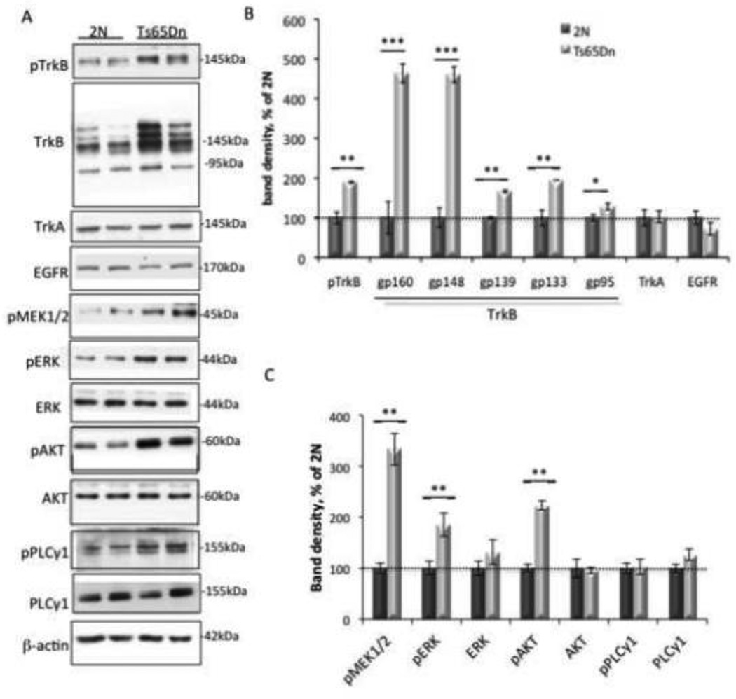
Figure 2. Excessive TrkB signaling in Ts65Dn synaptosomes.
(A) Representative WB of cortical synaptosomal lysates immunoblotted with antibodies against the tyrosine-phosphorylated receptors TrkB, TrkA, EGFR, MAPK, PI3K, PLCγ-related signaling proteins, and β-actin as a control for total protein levels. (B and C) Quantitative analysis of WB data showing a significant increase in total TrkB, pTrkB, pMEK1/2, pERK, and pAKT in Ts65Dn synaptosomes. Data are expressed as mean ± SEM percent of 2N values, n = 6 per genotype. *p<0.05, **p<0.005, ***p<0.0005, versus 2N.

