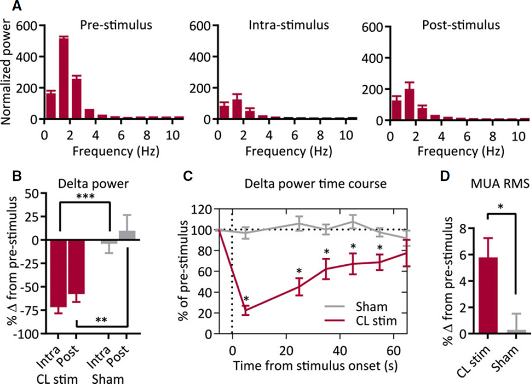
Figure 2.
Group data for thalamic CL stimulation under anesthesia. (A) Thalamic CL stimulation under anesthesia decreases low-frequency frontal cortical power during intrastimulus and poststimulus epochs; n = 12 animals. (B) Delta-band (0–4 Hz) power significantly decreases during intrastimulus and poststimulus epochs as compared to sham stimulation. (C) Time course of decreases in delta-band power is sustained for >50 s after 20 s stimulation. Stimulus-onset time = 0 s. (D) Decrease in low-frequency power is associated with a significant increase in MUA; n = 12 animals. All results are mean ± standard error of the mean (SEM). MUA, multiunit activity; RMS, root-mean-square amplitude; *p < 0.05, **p < 0.01, ***p < 0.001. Comparisons are two-tailed Student’s t-test.
Epilepsia © ILAE

