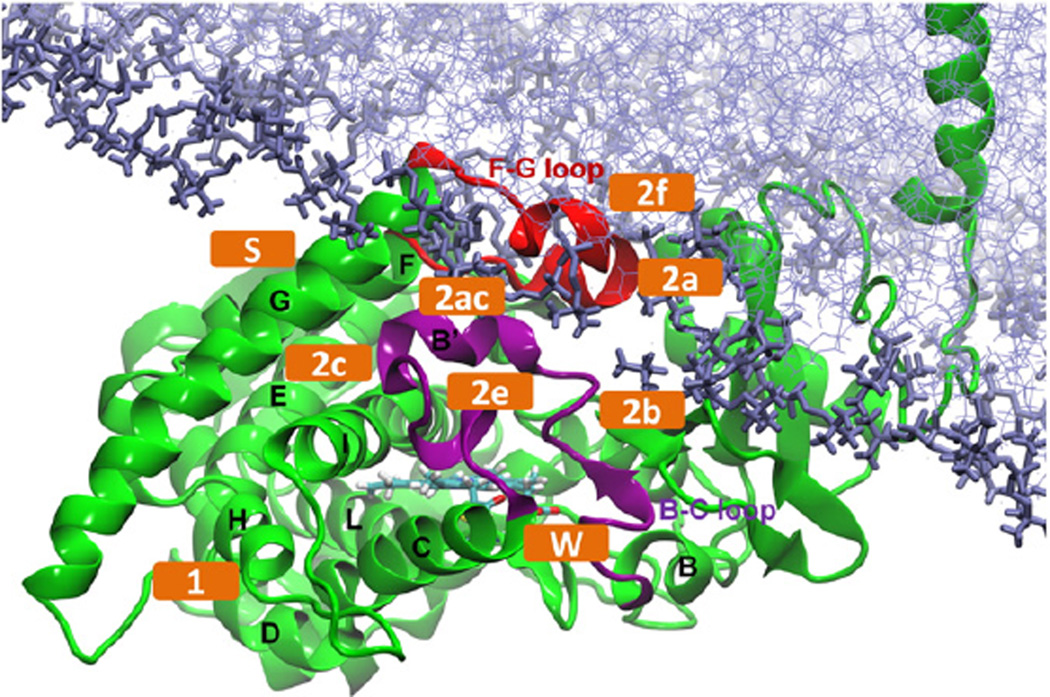
Fig. 1.
Model of membrane-bound ligand-free T. brucei CYP51 [8]. The important tunnels defined in Wade's nomenclature are labeled (orange). The protein is shown in cartoon representation with the B-C loop colored purple, the F-G loop red and the helices labelled. The heme (cyan) and the POPC bilayer (purple) are shown in stick representation.

