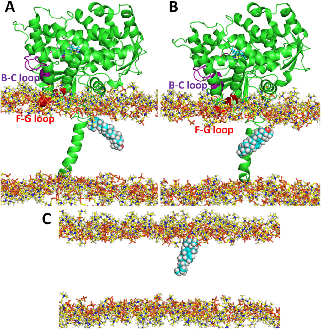
Fig. 7.
The change in orientation of OBT_DM in the membrane upon exit from T. brucei CYP51. (A) At the end of a RAMD simulation of T. brucei CYP51, the OBT_DM was orientated with its hydrophilic head down in the middle of the bilayer. (B) After a subsequent standard MD simulation of 6.5 ns, OBT_DM had changed its orientation in the membrane by about 100° so that its hydrophilic head pointed up towards the lipid head groups. (C) A further subsequent simulation without the protein starting with the snapshot of (B) allowed the hydrophilic part of OBT_DM to orient in the head group regions of the phospholipid bilayer. Only the head groups of the POPC bilayer are shown for clarity. OBT_DM is shown in CPK representation and T. brucei CYP51 is shown in cartoon representation.

