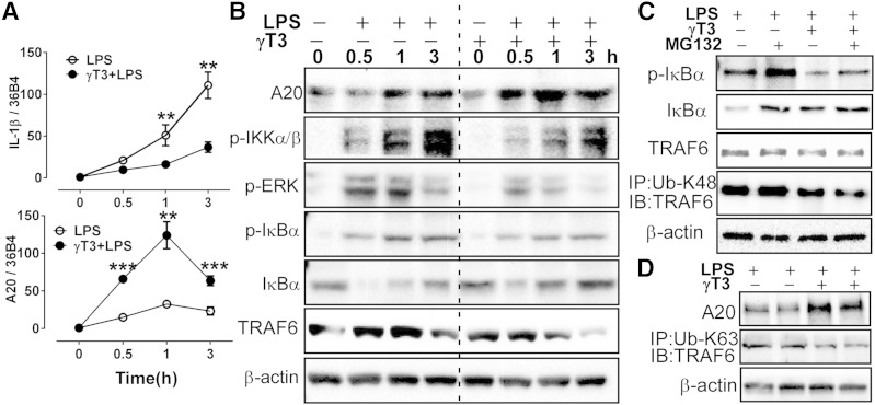
Fig. 6.
γT3 blocked NFκB-mediated priming of the inflammasome in BMDMs. BMDMs were pretreated with vehicle (−) or γT3 (+) for 24 h. A, B: Cells were stimulated with LPS (100 ng/ml) at t = 0 h, then total RNA and cellular proteins were extracted at 0, 0.5, 1, and 3 h. A: Temporal changes in mRNA levels of Il-1β and A20 by qPCR. B: Temporal changes in protein abundance of A20, p-IKK, p-ERK, p-IκBα, IκBα, TRAF6, and β-actin by Western blot analysis. C: BMDMs were pretreated with proteasomal degradation inhibitor MG132 (50 μM) prior to LPS stimulation (1 h). Protein levels of p-IκBα, IκBα, TRAF6, and β-actin were determined by Western blot. Cell extracts were immunoprecipitated (IP) with K48 ubiquitination antibody (Ub-K48) then immunoblotted (IB) with antibodies against TRAF6. D: BMDMs were stimulated with LPS for 1 h. Cell extracts were immunoprecipitated (IP) with K63 ubiquitination antibody (Ub-K63) then immunoblotted (IB) with antibodies against TRAF6. Data are shown as mean ± SEM. ** P < 0.01; *** P < 0.001.

