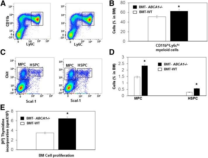
Fig. 6.
Hematopoietic ABCA1 deletion led to myeloid hyperplasia and hyper-HSPC proliferation with HFHSC diet feeding. Analysis of BM cells by flow cytometry of BMT-WT and BMT-ABCA1−/− mice fed the HFHSC diet for 21 weeks. A: Representative histogram of myeloid cell in BM. B: Myeloid cells were defined as CD11bhi and Ly6chi and quantified in BM. C: Representative histogram of MPCs and HSPCs in BM. D: MPCs were identified as Sca1− and ckit+ and HSPCs were identified as Sac1+ and ckit+ and were quantified and shown as percentage of total BM cells. E: BM was isolated from HFHSC diet-fed BMT-WT and BMT-ABCA1−/− mice and incubated with IL-3 and GM-CSF, and proliferation was assessed by [H3]thymidine incorporation. Results are means ± SD of five mice per group. *P < 0.05 versus BMT-WT.

