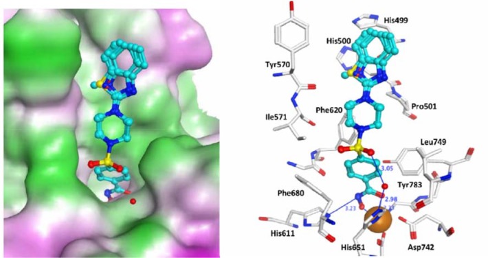
Fig 8. Docking poses of compounds 9a-d in the HDAC6 catalytic domain.
Inhibitors are colored in cyan. The zinc ion is shown as a brown ball. Hydrogen bonds between HDAC6 and inhibitors are shown as blue lines and distances are given in Å. On the right side the molecular surface is displayed and contoured according to the hydrophobic potential (magenta = hydrophilic, green = hydrophobic).

