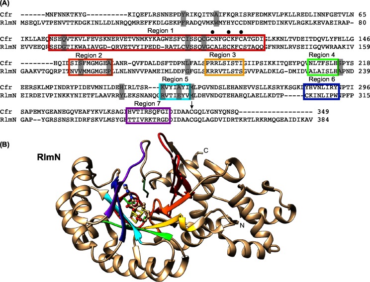
Fig 3. The interchanged regions for the investigation of C2/C8 specificity.
(A) Alignment of the amino acid sequences of Cfr (GI: 34328031 / NCBI: NP_899167.1) and RlmN (GI: 16130442 / NCBI: NP_417012.1). The seven coloured boxes depict the regions that encompass the active site of the enzymes. The dots above the sequence indicate the CX3CX2C motif. The black arrow shows the cysteine 338/355. The grey shading mark the 13 selectively conserved positions in Cfr- or RlmN-like proteins. (B) Representation of the X-ray crystal structure of RlmN (PDB file 3RFA) [26] with the same region colouring as in the amino acid alignment and oriented similar to the Cfr model structure in Fig 2A. Also, the SAM molecule and the [4Fe-4S] cluster are included as in Fig 1A. The three cysteines in the CX3CX2C as well as the Cys355 are shown in green sticks.

