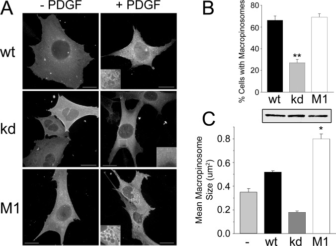
Fig 2. Rescue of PDGF-induced macropinocytosis.
DGKζ-null cells were infected with adenoviruses bearing HA-tagged versions of wild type (wt), kinase dead (kd), or phosphomimetic (M1) DGKζ. (A) Representative images of infected cells with (+ PDGF) and without (- PDGF) stimulation. Insets show magnified images of the regions indicated by the white boxes. Scale bars, 20 um. (B—C) Quantification of macropinocytosis in infected DGKζ-null cells. The graphs show the percentage of cells with macropinosomes (B) and the mean macropinosome size (C). Values are the mean ± SEM from three independent experiments. Asterisks denote a significant difference (p<0.05) from kd as determined by a one-way analysis of variance, followed by a Tukey post hoc multiple-comparison test. The western blot shows equivalent levels of HA-DGKζ expression in lysates of infected cells. The lanes correspond to the bars in the graphs above and below.

