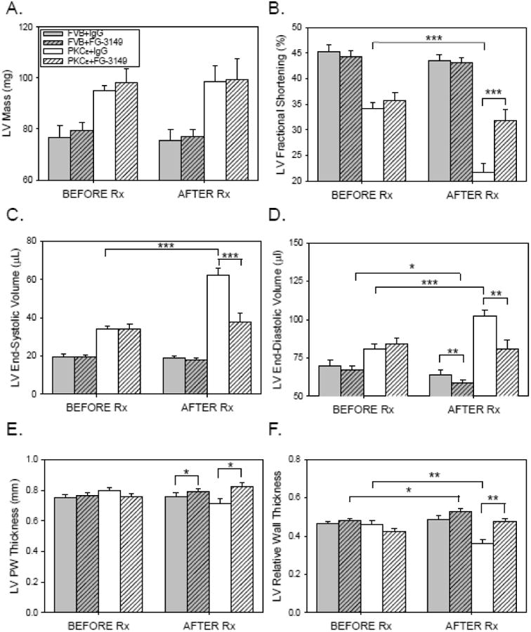
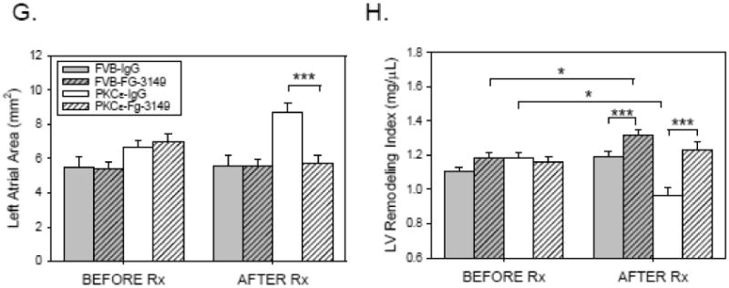
Figure 2. CTGF neutralizing antibody maintains LV function and slows the progression of LV remodeling in PKCε mice.


3 month-old FVB (n=22; filled bars) and PKCε (n=22; open bars) mice underwent baseline M-mode and 2-D echocardiography (BEFORE Rx), and were then randomly assigned to receive nonimmune mouse IgG (IgG; 30mg/kg IP; no cross-hatch) or FG-3149 (30mg/kg IP; cross-hatched), a neutralizing mAb to CTGF. Animals were treated twice weekly for a period of 3 months, and were then subjected to repeat M-mode and 2-D echocardiography (AFTER Rx). (A) LV Mass (mg); (B) LV Fractional Shortening (%); (C) LV End-systolic Volume (μL); (D) LV End-diastolic Volume (μL); (E) LV Posterior Wall (PW) Thickness (mm); (F) LV Relative Wall Thickness; (G) Left Atrial Area (mm2); and (H) LV Remodeling Index (mg/μl). Data are means±SEM for n=10-12 mice in each treatment group. Data from the 4 groups BEFORE and AFTER Rx were compared by 2-way ANOVA followed by the Holm-Sidak test. Data for each group BEFORE and AFTER Rx were compared by paired t-test. P<0.05 (*), 0.01 (**), or 0.001 (***).
