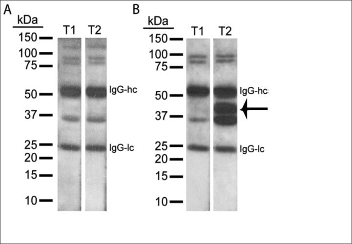
Figure 1.
Western blots of human SCI plasma Time 1 and Time 2. Pictures of representative western blots probed with T1 (<48 hr) and T2 (2-4 weeks post-injury) plasma from two SCI patients. A. Although a few cross-reacting bands are detected for this patient, the intensity of immunoreactive bands was not specifically enhanced at T2. B. T2 plasma from a second patient cross-reacted with bands ranging from 36 to 42 kDa whose intensities were higher compared to T1 plasma from the same patient. (IgG-hc: Immunoglobulin G heavy chain, IgG-lc: Immunoglobulin G light chain.)

