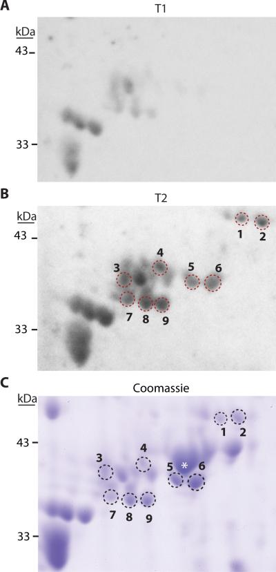
Figure 2.
Western blots and Coomassie stained gel showing increased immunoreactivity after SCI. Picture of a representative western blot from membranes probed with A. T1 plasma, and B. T2 plasma from a patient positive for autoantibodies. C. Coomassie-stained 2D gel from the same area as shown in the membranes. Spots excised for LC-MS/MS are indicated by numbers; * indicates location of β-actin. (T1, (<48hrs) T2, (2-4 weeks) post-injury.)

