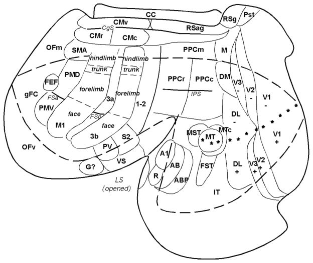
Fig. 1.
A surface view of the flattened neocortex of a prosimian primate (Galago garnetti). Cortex has been separated from the rest of the brain and flattened as a single sheet with proposed cortical areas imposed. This view shows cortex normally hidden in views of the intact brain. The dashed lines roughly outline the cortex that would be visible on a dorsolateral view of the cerebral hemisphere. Posterior parietal cortex contains rostral (PPCr), caudal (PPCc) and medial (PPCm) regions. Primary (V1) and secondary (V2) visual areas are common to most mammals. As in other primates galagos also have the third visual area (V3), a dorsomedial area (DM), a middle temporal area (MT), a dorsolateral area (DL or V4), fundal area of the superior temporal sulcus (FST), a MT crescent (MTc), and inferior temporal cortex (IT) of several divisions. Auditory cortex includes A1 and rostral (R) areas, plus presumed auditory belt (AB) and parabelt (APB) regions. Somatosensory cortex includes a primary area (S1 or area 3b) (note the overall somatotopy), a proprioceptive area 3a and secondary areas of 1–2, S2, parietal ventral (PV) and ventral somatosensory (VS). Motor areas include primary motor area (M1) ventral (PMV) and dorsal (PMD) premotor areas, supplementary motor area (SMA), frontal eye field (FEF) and ventral (CMv), rostral (CMr) and caudal (CMc) cingulate areas. Frontal cortex includes frontal granular cortex (gFC), medial (OFm) and ventral (OFv) orbital frontal regions. Agranular (RSag) and granular (Rsg) retrosplenial areas, area prostriata (Pst) are noted. CC= corpus callosum. Stars mark horizontal meridian. Modified from Kaas and Preuss, 2014.

