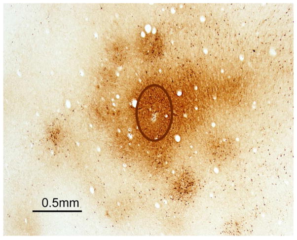
Fig. 4.
Dense connections of a reach domain in PPCr of a galago with surrounding satellites. The photomicrograph is of a brain section cut parallel to the cortical surface after an injection of a tracer (BDA) into the reach domain identified by electrical stimulation. The injection core (outlined oval) labeled neurons and axons most densely and uniformly within the domain and in a patchy pattern along the margins of the domain, and just outside the domain. Modified from Stepniewska et al., 2015.

