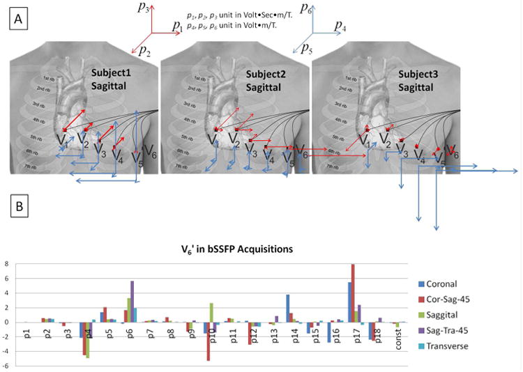
Figure 7.

(A) The first 6 fitted parameters (1st-order coefficients) for 3 subjects during sagittal bSSFP imaging with phase-encoding performed along the y direction (Anterior-Posterior). The 3D vector plots in the center illustrate graphically the size and direction of the coefficients for the precordial electrodes V1'-V6', with the origin of each vector placed at the approximate spatial position of the respective electrode. Red arrows denote p1k – p3k, which multiply the gradient derivatives along the 3 directions, and blue arrows denote p4k – p6k, which multiply the 3 gradients. A gradually increasing influence of the magnetic gradient fields on the ECG noise was observed from V1' to V6' in all subjects, although the magnitude of the gradient-induced voltage varied greatly among subjects. (B) Variation of the 19 fit coefficients during bSSFP acquisitions along different slice planes in a volunteer. Cor-Sag-45 and Sag-Tra-45 designate 45-degree oblique angles between the Coronal and Sagittal, and Sagittal and Transverse orientations, respectively.
