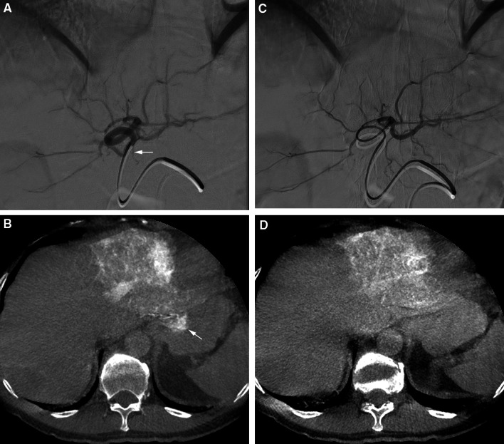
Fig. 4.
A DSA from the LHA. B C-arm CT performed from the LHA shows extrahepatic shunting in the gastric wall (white arrow). The small extrahepatic branch indicated by the white arrow in (A) was the culprit vessel. C The catheter was positioned more distal in the LHA. D C-arm CT performed from the new injection position did not show extrahepatic shunting anymore

