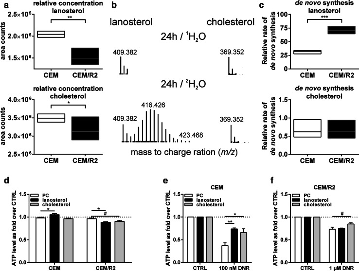
Fig. 2.
Resistant leukemia cells CEM/R2 exhibit an increased flux through the lanosterol but not the cholesterol pool and are negatively affected by exogenous lanosterol addition. a Relative concentration of lanosterol and cholesterol in CEM versus CEM/R2 cells as determined by LC–MS. b Mass spectra of lanosterol (left) and cholesterol (right) after cell growth for 24 h using regular media (top) and media with addition of 30 % 2H2O (bottom). c Data showing de novo synthesis of lanosterol and cholesterol measured on cells grown in 30 % 2H2O. a–c Data of a single experiment carried out in five replicates are shown as minimum to maximum with line at mean. d Viability of CEM and CEM/R2 cells (n = 4) that were grown for 48 h in serum-free RPMI 1640 in presence of 50 μM 1,2-dimyristoyl-sn-glycero-3-phosphocholine (PC), lanosterol/PC mixture (each 25 μM), or cholesterol/PC mixture (each 25 μM). Viability of CEM (e) and CEM/R2 (f) cells that were incubated for 48 h in absence or presence of DNR (CEM, 100 nM and CEM/R2, 1 μM) and 50 pM PC, lanosterol/PC mixture (each 25 μM), or cholesterol/PC mixture (each 25 μM). d–f Data are shown as mean ± SEM of four independent experiments carried out in triplicate, P values were determined using an ordinary one-way ANOVA with Dunnett’s multiple comparisons test. #≤0.1; *P ≤ 0.05; **P ≤ 0.0110

