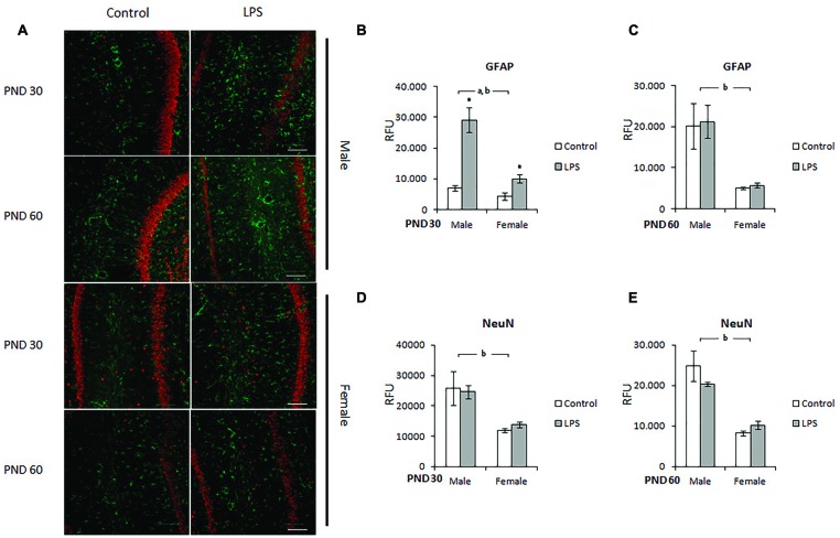
FIGURE 4.
Immunohistochemistry for GFAP and NeuN in the CA1 hippocampus of LPS-offspring rats. (A) Shows immunohistochemistry for GFAP (green) and NeuN (red) in hipocampal slices of 30- and 60-day old Wistar rats prenatally exposed to LPS (males and females). GFAP and NeuN were then quantified in the hippocampal slices of PND 30 (B,D, respectively) and PND 60 Wistar rats prenatally exposed to LPS (C,E, respectively). Data are expressed as means ± standard error (LPS group, N = 5; control group, N = 5); the experiments were performed in triplicate. aSignificant effect of prenatal treatment; bsignificant effect of sex (Two-way ANOVA, p < 0.05). ∗Significantly different from control (Bonferroni’s post hoc, p < 0.05). Scale bar = 10 mm.

