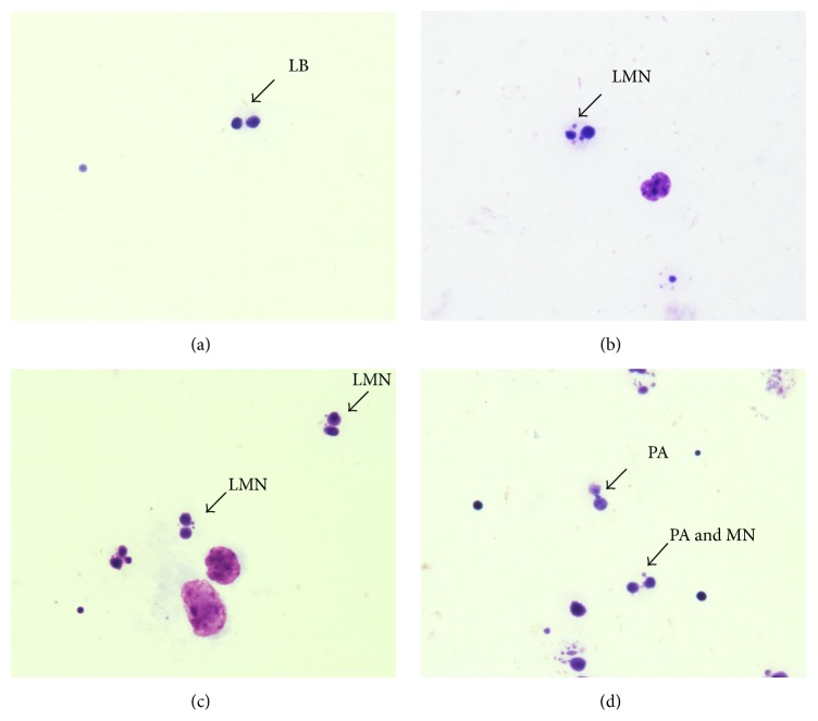
Figure 3.
(a) Photomicroscopy of binucleated lymphocyte without micronucleus (LB), observed in negative control. Image of binucleated lymphocyte with micronucleus (LMN), observed in positive control (b) and group treated with E6 recombinant oncoprotein (c). Image of binucleated lymphocyte with anaphase bridge and micronucleus (PA and MN), observed in group treated with E6 (d). Images obtained with total magnification of 1,000x.

