Figure 5.

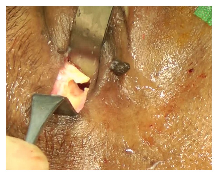
The periost layer was elevated exposing the bone at the sutura frontozygomatica. At this point an L-shaped canal was drilled, providing flexible stability and optimizing angulation for the cable entering the orbit.

The periost layer was elevated exposing the bone at the sutura frontozygomatica. At this point an L-shaped canal was drilled, providing flexible stability and optimizing angulation for the cable entering the orbit.