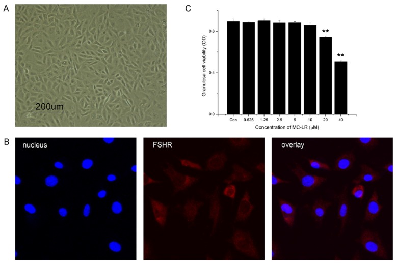
Figure 5.
MC-LR affects cell viability of mGCs. (A) Primary mGCs were obtained from mouse ovary and an optical microscope image was taken; (B) mGCs cultured on coverslips were fixed and immunostained with specific antibodies for FSHR; (C) effects of 48 h exposure to MC-LR on mGCs cell viability. mGCs were treated with 0, 0.625, 1.25, 2.5, 5, 10, 20 and 40 μM MC-LR, respectively. 48 h later, measurement of cell viability was carried out with CCK-8. The percentage of dead cells was increased in a dose-dependent manner. The analysis were performed with three replications and representative data was shown. (** p < 0.01).

