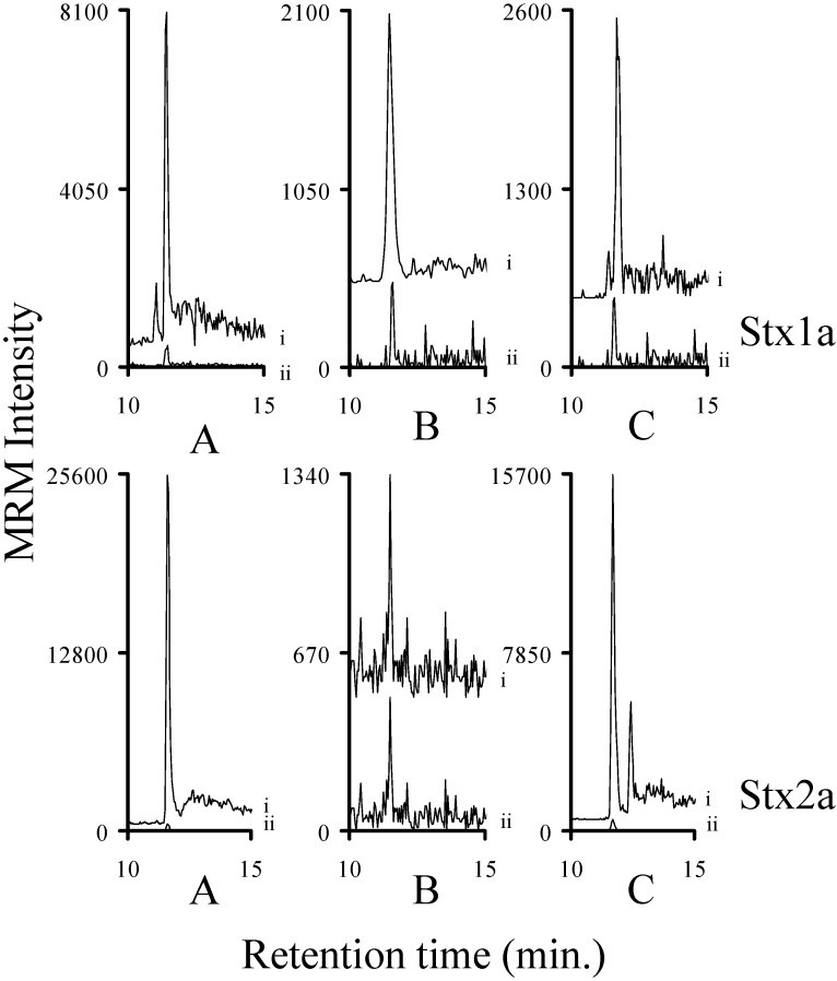
Figure 6.
The intensity of the MRM signals derived from the y8 ion of the analyte decapeptide of Stx1a or Stx2a (i) in sterile filtered bacterial medium (A); human serum (B); and human serum denatured with guanidinium chloride (C). The corresponding 15N-labeled internal standard is normalized to 500 (ii) to allow the signal from the analyte decapeptide to be viewed on the same plot. The amount of Stx1a in each injection (n = 2) was calculated to be (left (l) to right (r)) 5.8 ± 0.4 fmol, 3 ± 1 fmol, and 1.7 ± 0.8 fmol, respectively. The amount of Stx2a in each injection (n = 2) was calculated to be (l to r) 29 ± 0.4 fmol, 0.08 ± 0.05 fmol, and 10 ± 0.6 fmol, respectively.

