Figure 1.
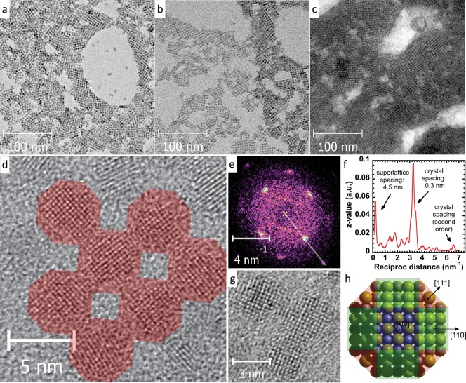
(a–c) TEM micrographs of PbS CQD solids formed via exposure to (a) NH4I, (b) MAI, and (c) TBAI; (d, e) real-space and Fourier-transformed HRTEM images, and (f) an extracted profile from the FT image of a square domain showing that the superlattice consists of CQDs oriented the same direction after treatment with MAI; (g) high-resolution image on the oriented assembly showing epitaxially connected CQDs; (h) schematic structure with the main facets of a PbS CQD.

