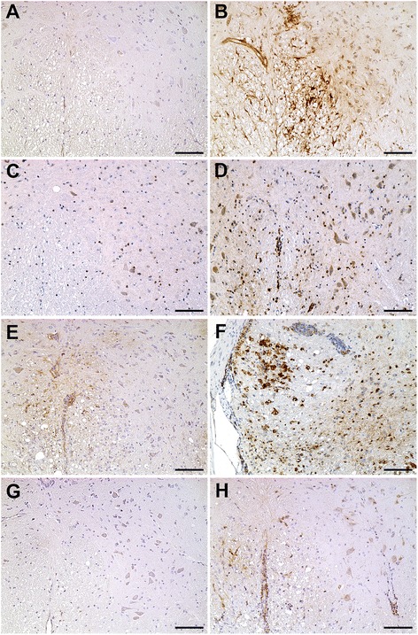
Fig. 3.

ISG15, PKR, and OAS1 protein expression in the spinal cord of mock- and TMEV-infected SJL/J mice. a Low expression of ISG15 proteins in mock-infected mice at 42 dpi. b High expression in endothelial cells and astrocytes of TMEV-infected SJL/J mice at 42 dpi. c Few cells express PKR proteins in mock-infected SJL/J mice at 196 dpi. d Many oligodendrocytes, microglia/macrophages, and neurons express PKR proteins in TMEV-infected SJL/J mice at 196 dpi. e Few intralesional cells express ISG15 at 98 dpi. f High PKR expression in intralesional gitter cells and some perivascular immune cells in TMEV-infected SJL/J mice at 98 dpi. g Very low expression of OAS1 proteins of mock-infected SJL/J mice at 98 dpi. h Few neurons, microglia, and perivascular immune cells express OAS1 proteins in TMEV-infected SJL/J mice at 98 dpi. Immunohistochemistry visualized by the avidin-biotin-peroxidase complex method with 3,3-diaminobenzidine as substrate and counterstaining with Mayer’s hematoxylin. Bar 100 μm
