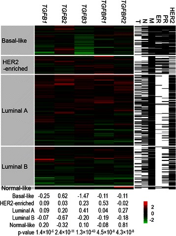
Fig. 3.

Heatmap visualization of tumour mRNA levels. Tumour samples from the TCGA cohort were hierarchically clustered according to the log2-transformed and standardized mRNA expression levels of TGFB1, TGFB2, TGFB3, TGFBR1 and TGFBR2 for each PAM50 subtype in the middle panel, where each row represented a tumour specimen and each column represented a gene. The colour represented the mRNA expression levels (green: low, red: high). The median mRNA levels of each gene for each PAM50 subtype were shown at the bottom, and the p-values were calculated from the Kruskal-Wallis test for differences in the mRNA levels across the PAM50 subtypes. All the p-values were still significant after Bonferroni correction for multiple testing. The tumour diameter (T), lymph node metastasis (N), distant metastasis (M), ER PR and HER2 statuses of each specimen were shown in the right-side columns, white: positive or >2 cm, black: negative or ≤2 cm, grey: missing data
