Fig. 1.

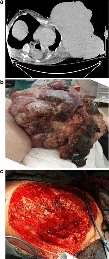
Gross appearance of the mass at presentation (a, left panel), computed tomography image without contrast of patient at presentation showing large exophytic mass (b, center panel), and postoperative appearance (c, right panel)

Gross appearance of the mass at presentation (a, left panel), computed tomography image without contrast of patient at presentation showing large exophytic mass (b, center panel), and postoperative appearance (c, right panel)