Fig. 4.

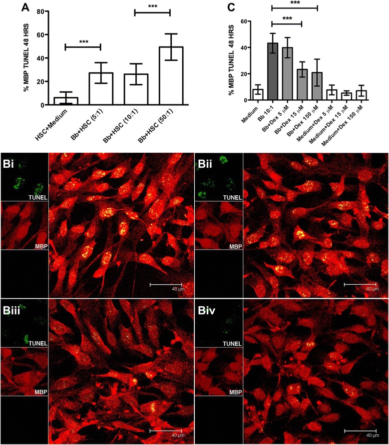
Dexamethasone protects HSC from Bb-induced apoptosis in a dose-dependent fashion. A Graphical representation of the percent apoptosis of HSC as induced by increasing MOI of live Bb. Representative images of the quantitative view of apoptosis after immunofluorescence staining and visualized by confocal microscopy by the in situ TUNEL assay (green) as induced by live Bb (MOI 10:1) in HSC stained with MBP (red), after 48 h, (Bi), Bb in the presence of 5 μM dexamethasone (Bii), 15 μM dexamethasone (Biii), and 150 μM dexamethasone (Biv), respectively. C Graphical representation of the effect of dexamethasone on the levels of apoptosis in HSC as induced by live Bb as visualized by the in situ TUNEL assay. The one-way ANOVA and Tukey’s multiple comparison test was used to evaluate the statistical significance between means and SD of ten data sets (approximately 500 cells) for each condition, ***p < 0.001
