
Figure 1. The neural circuitry of mood.
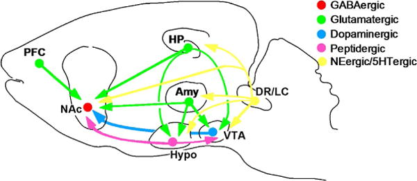
The figure shows a highly simplified summary of a series of neural circuits in the brain that are believed to contribute to the regulation of mood. While most research in the depression field until recently has focused on hippo-campus (HP) and cerebral cortex (e.g., prefrontal cortex or PFC), there is the increasing realization that several subcortical structures implicated in reward, fear, and motivation are also critically involved. These include the nucleus accumbens (NAc), amygdala (Amy), and hypothalamus (Hypo). The figure shows only a subset of the many known interconnections among these various brain regions. The figure also shows the innervation of several of these brain regions by monoaminergic neurons. The ventral teg-mental area (VTA) provides dopaminergic input to the NAc; inputs to most of the other brain areas are not shown in the figure. Norepinephrine (NE, from the locus coeruleus or LC) and serotonin (5HT from the dorsal raphe and other raphe nuclei) innervate all of the regions shown in the figure. In addition, strong connections between the hypothalamus and VTA–NAc pathway have been established in recent years. From Nestler and Carlezon (2006) with permission.