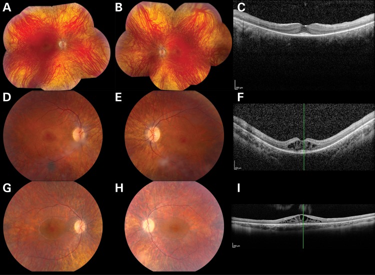
Figure 2.
Color fundus photographs (A, B, D, E, G, H) and OCT (C, F, I) from individuals P1 (A–C), P2 (D–F) and P3 (G–I) affected with TRNT1-associated autosomal-recessive RP. The color photographs reveal vascular narrowing in all three individuals, subtle bone spicule-like pigmentation in P1 and cystoid macular edema in P2 and P3. OCT reveals thinning of the photoreceptor layers in all three individuals and marked cystoid macular edema in P2 and P3.

