Figure 5.
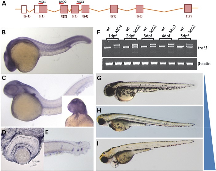
trnt1 expression and knockdown in zebrafish embryos. (A) Zebrafish trnt1 gene structure noting the location of MOs used for knockdown. (B) Zebrafish trnt1 transcript is broadly expressed throughout the embryo up to 1 dpf. (C) By 2 dpf, trnt1 becomes enriched in the brain, pectoral fin, blood and heart (inset). (D) Section through the central retina of 3 dpf larva showing trnt1 expression. (E) trnt1 expression in blood forming regions and the neuromast. (F) RT-PCR of RNA isolated from control and morphant embryos showing altered splicing in MO-injected embryos through 5 dpf. Uninjected WT embryos are used as control. Phenotypic range of morphological defects observed in trnt1 morphants at 5 dpf. (G) WT-like, (H) reduced eye size and blood flow and (I) eye and cardiovascular defects. Blue bar represents the penetrance of morphological defects with MO dose: 6.5 ng generating 80% defective, 4.5 ng generating 60% defective and 1.5 ng inducing defects in 15% of injected embryos.

