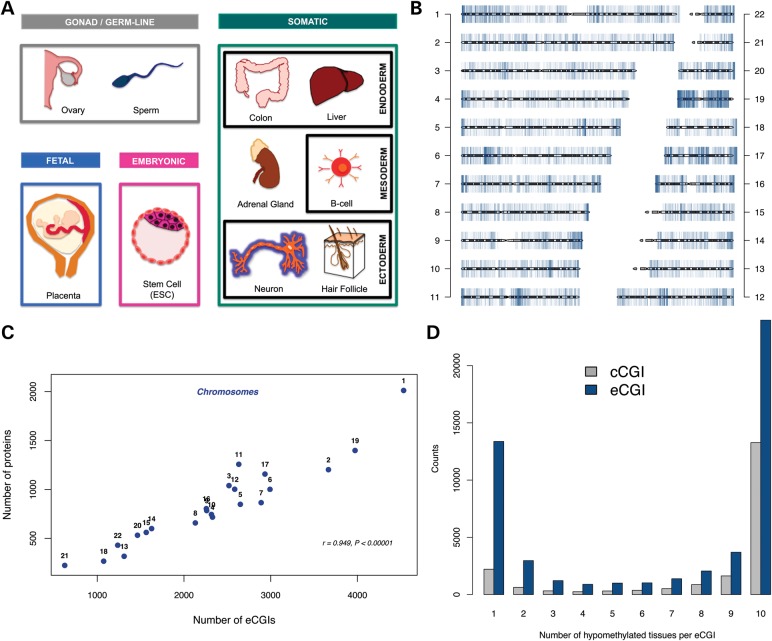
Figure 1.
(A) Tissues analyzed for eCGI identification, including embryonic, gonad, germ line and fetal tissues, as well as six adult somatic tissues of distinct developmental origins. These were selected to have the highest cell type diversity with respect to gene expression patterns (68) while avoiding overly cell heterogeneous tissues. Ovaries comprise germ-line cells and endoderm-derived tissue. The adrenal gland has both ectodermal (medulla) and mesodermal (cortex) origins. (B) The genomic distribution of eCGIs. (C) The correlation between the numbers of protein-coding genes and eCGIs on each chromosome. (D). Distribution of eCGIs and cCGIs across tissues.

