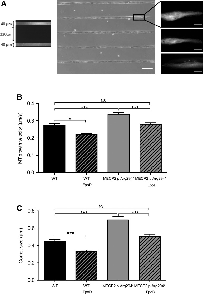
Figure 3.
Human MECP2 p.Arg294* astrocytes present a higher MT growth velocity and a higher EB3-comet size than wild-type astrocytes in standardized morphology condition, that can both be corrected by EpoD. (A) Cells were seeded on PLL-PEG-coated coverslips printed with 40 µm large lines of fibronectin and presented subsequently a homogeneous polarized morphology. Fluorescent fibronectin lines are shown in the left panel. The middle panel illustrates the cell alignment on fibronectin lines (white light) and the right panel gives examples representative of the homogeneous morphology adopted by the EB3-GFP expressing cells prior to time-lapse microscopy. (B and C) EB3-GFP transfected wild-type (WT) and MECP2 p.Arg294* isogenic astrocytes were incubated with 10 nm EpoD or vehicle for 1 h before timelapse imaging. EB3-GFP movement corresponding to MTs end growing radially from the centrosome region was followed in single cells by time-lapse microscopy (image acquisition every 1 s during 1 min, 61 images) and EB3-GFP velocity and comet size were measured. A total of 28 WT, 46 MECP2 p.Arg294* vehicle-treated cells, 8 WT and 36 MECP2 p.Arg294* EpoD-treated cells were included in the analysis. Bars represent (B) mean EB3 comet velocity and (C) mean EB3 comet size, ± SEM. *A P-value for Student's test under 0.05, ***under 0.001 and NS over 0.05. White scale bar represents 200 µm, gray scale bar represents 10 µm.

