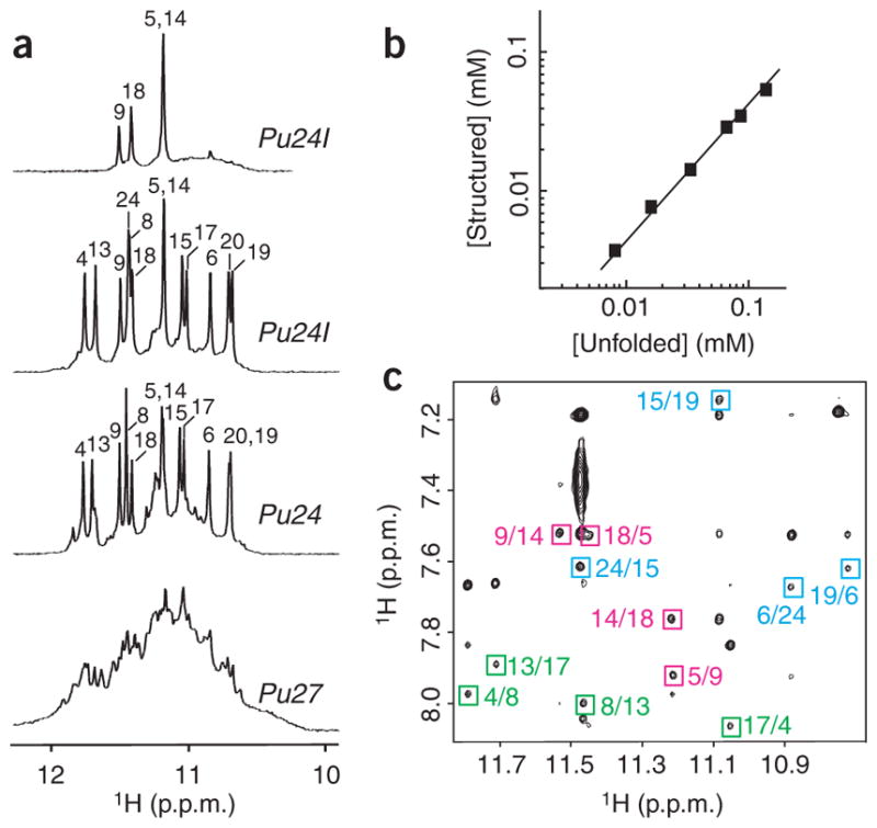
Figure 1.

NMR study of the MYC promoter guanine-rich sequences. (a) The 600 MHz imino proton spectra of Pu27 (bottom), Pu24 (lower middle) and Pu24I in H2O (upper middle) and of Pu24I after 4 h in D2O (top), with unambiguous resonance assignments for both Pu24 and Pu24I listed over the spectra. (b) Determination of stoichiometry of Pu24I by NMR (see Methods). Line of slope 1 is drawn through the data points. (c) NOESY spectrum (mixing time, 200 ms) of Pu24I. Characteristic imino-H8 cross peaks that identify three G-tetrads (colored cyan, magenta and green) are framed and labeled with the number of imino protons in the first position and that of H8 in the second position.
