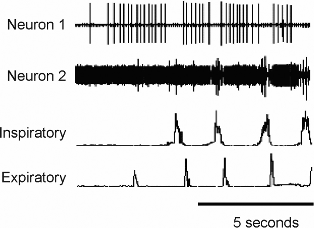
Figure 2. Activity patterns of putative E and I cough phasic neurons in the NTS.
Neuron 1 (a cough expiratory neuron) was silent during breathing and recruited before coughing began and neuron 2 is the large action potential that is recruited during the cough I phase. The recording for neuron 2 also has a smaller amplitude neuron that is tonically active but is transiently depressed during cough. Inspiratory-parasternal muscle EMG, Expiratory-transversus abdominis muscle EMG. Cough was induced by mechanical stimulation of the intrathoracic trachea in an anesthetized cat. Extracellular action potentials of NTS neurons were recorded with tungsten microelectrodes.

