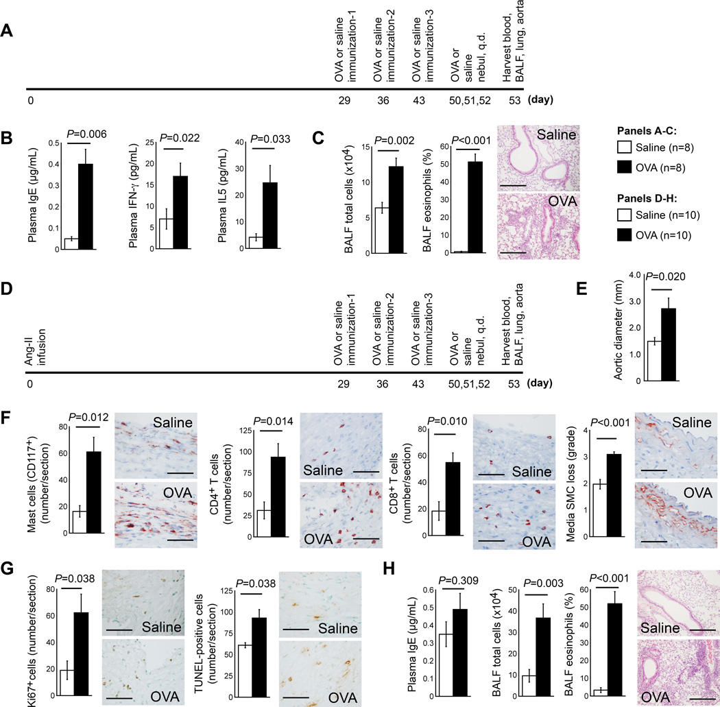
Figure 3.
OVA-induced ALI promotes progression of pre-established AAA in Apoe−/− mice. A. Experimental protocol of producing ALI alone. B. Plasma levels of IgE, IFN-γ and IL5. C. BALF total inflammatory cell number and eosinophil percentage. D. Experimental protocol of AAA production, followed by ALI production. E. Aortic diameters at harvest. F. AAA lesion contents of macrophages, CD4+ and CD8+ T cells, and media SMC loss in grade. G. AAA lesion numbers of Ki67-positive proliferating cells and TUNEL-positive apoptotic cells. Representative data for panels F and G are shown to the right, Scale bar: 50 µm. H. BALF total inflammatory cell number and eosinophil percentage. Representative lung histology data (H&E staining) in panels C and H are shown to the right, scale bar: 200 µm.

