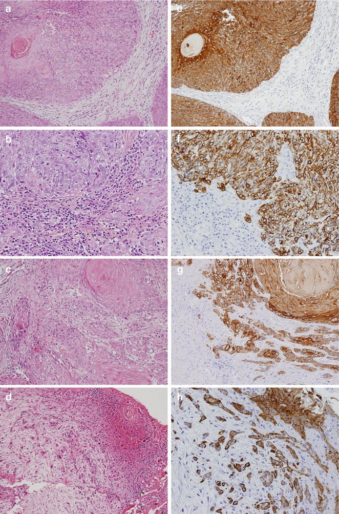
Fig. 1.

Hematoxylin-eosin staining of SCC of the EAC showing tumor budding/sprouting (a–d) and cytokeratin immunohistochemistry (CK-IHC) for tumor budding/sprouting (e–h) at the invasive front. Inset in a, e shows absence of tumor budding; budding grade 0, the others shows presence of tumor budding; b, f shows budding grade 1, c, g shows budding grade 2, and d, h shows budding grade 3
