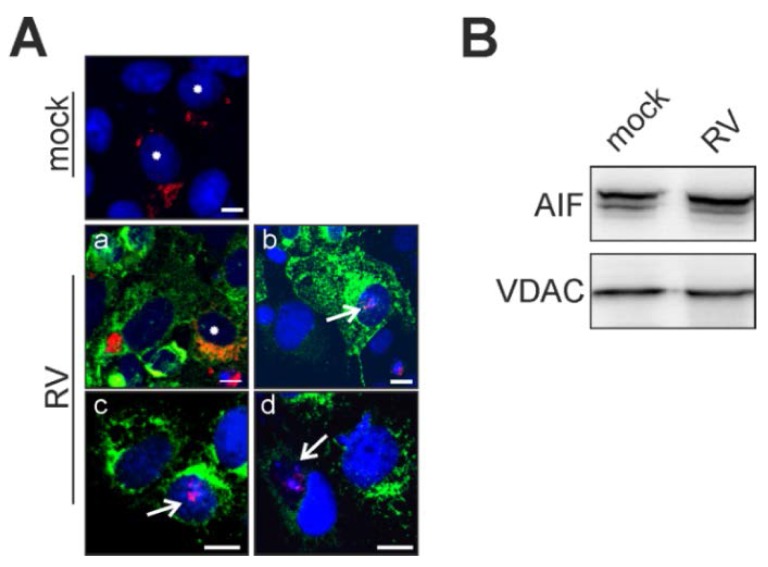
Figure 3.
Release of the mitochondrial death-effector protein AIF. Vero cells overexpressing AIF-TagRFP were applied at 2 dpi (A) for determination of RV-induced translocation of AIF-TagRFP (shown in red) from mitochondria to the nucleus. Nuclear DNA is shown in blue, the viral E1 protein is shown in green. Asterisks (mock and Aa) indicate physiological localization of AIF to mitochondria. Arrows (Ab,c and d) highlight cells with nuclear association of AIF; (B) 30 μg of mitochondrial fractions obtained at 3 dpi from mock- and RV-infected Vero cells were subjected to Western blot analysis with anti-AIF antibody. As a loading control, anti-VDAC antibody was applied. Scale bar, 10 μm.

