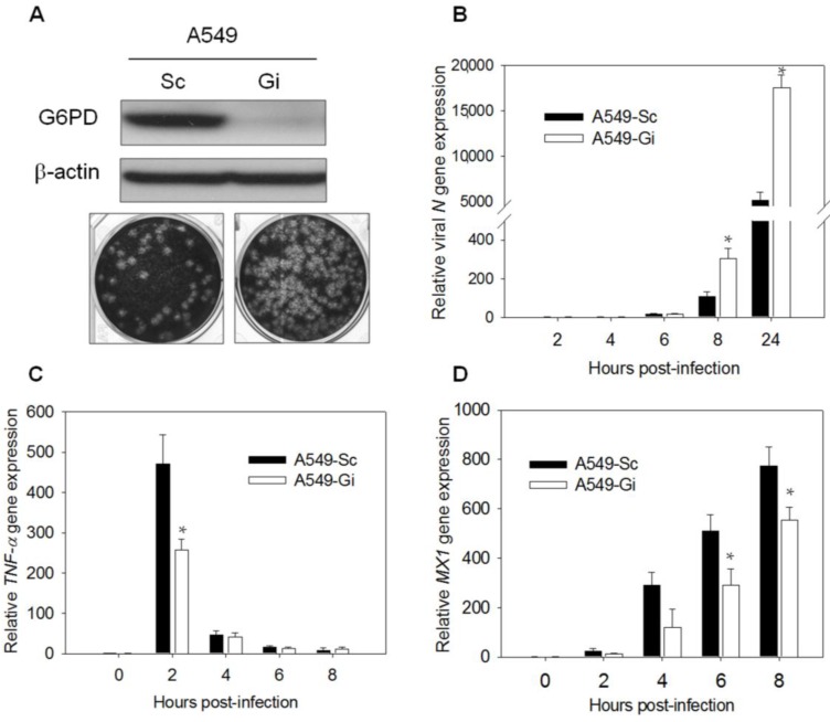
Figure 1.
Expressions of antiviral gene MX1 and TNF-α decrease upon HCoV 229E infection in A549-Gi cells. (A) A549-Sc and -Gi cells were harvested for determination of G6PD expression by western blotting. β-Actin was used as internal control. A549-Sc and -Gi cells were infected with HCoV-229E (0.1 MOI) for 24 h then viral particle was harvested and production was determined using plaque assay; (B) A549-Sc and -Gi cells were infected with HCoV-229E (0.1 MOI) for indicated time points. Viral N gene expression was determined by quantitative-PCR. Data were normalized to the value of infected A549-Sc cells at 2 h p.i.; (C) RNA was harvested from HCoV-229E-infected cells at indicated time p.i.. TNF-α gene expression was determined by quantitative-PCR. Data were normalized to the value of uninfected A549-Sc cells; (D) RNA was harvested from HCoV 229E-infected cells at indicated time points p.i.. MX1 gene expression was determined by quantitative-PCR. Data were normalized to the value of uninfected A549-Sc cells. Values represent average ± SD of three experiments. * p < 0.05 as compared to A549-Sc cells.

