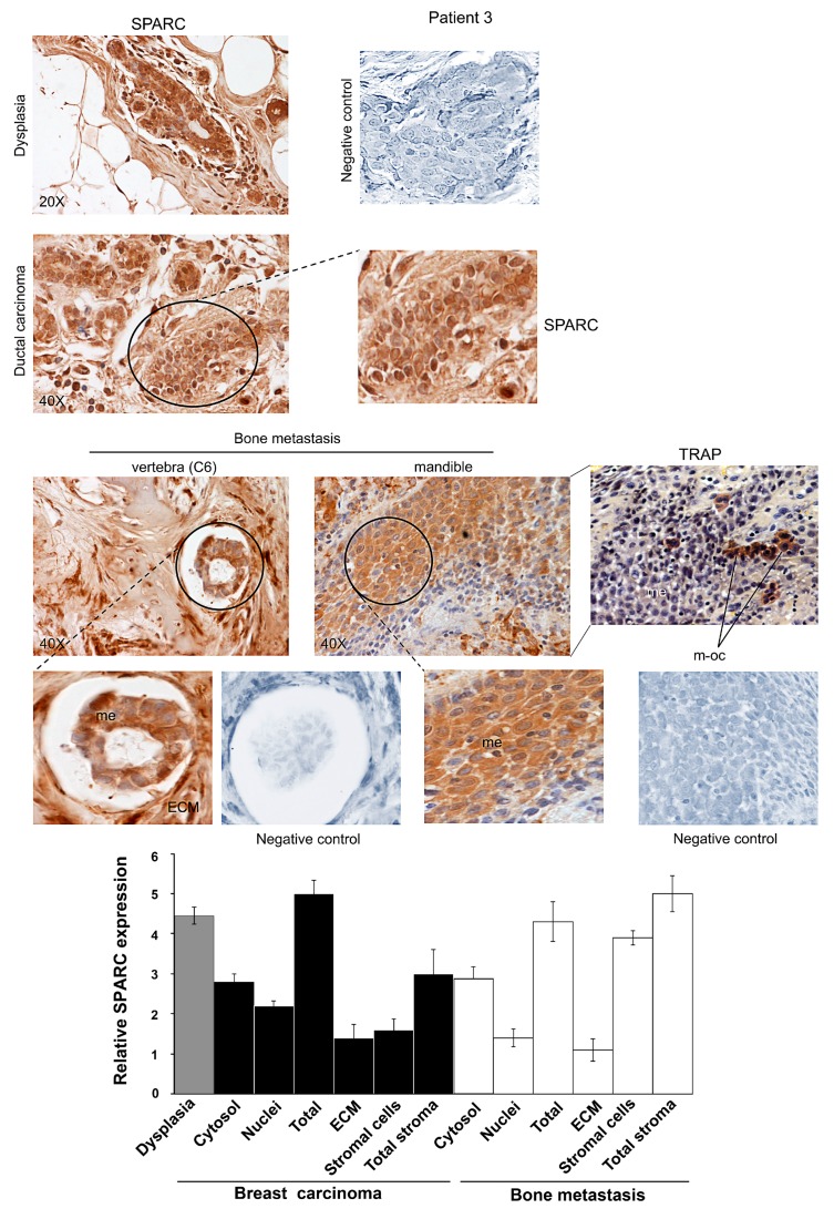
Figure 3.
SPARC expression during ductal breast carcinoma progression from dysplasia until bone metastasis for Patient 3. Representative images of immunohistochemical staining for SPARC were shown. In bone metastasis established in the mandible, TRAP assay was also performed. Magnifications of details are shown. ECM, Extracellular matrix; me, metastatic cells; m-oc, mature osteoclasts. Negative controls were assayed without the specific antibody. The histograms, showing the relative values for SPARC expression, were prepared by using the score values reported in Supplementary Table S1. The data are the means ± S.E. of experiments performed in triplicate.

