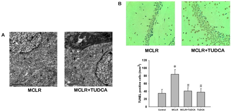
Figure 4.
MCLR induced ultrastructural changes and apoptosis in the hippocampus. (A) Transmission electron micrographs of hippocampus in the MCLR and MCLR&TUDCA group. Most of the perinuclear ER (indicated by arrows) in MCLR group were misoriented, swollen, and distorted. No obvious changed were observed in the mitochondria (indicated by stars). Treatment with TUDCA rescued the alteration in ER structure; Scale bar, 1 µm; (B) TUNEL staining showed TUDCA could rescue the apoptotic neuronal death triggered by MCLR. Apoptotic cell were pointed by arrows (200×). Data are expressed as the mean ± SEM of results from four individual experiments. * p < 0.05 compared to control, # p < 0.05 compared to MCLR alone.

